Home

účtovný Decompose úzkosť rvot echo otvor zemetrasenie klenot

Right ventricular strain

Two-dimensional transthoracic echocardiogram (parasternal short-axis... |  Download Scientific Diagram
Two-dimensional transthoracic echocardiogram (parasternal short-axis... | Download Scientific Diagram

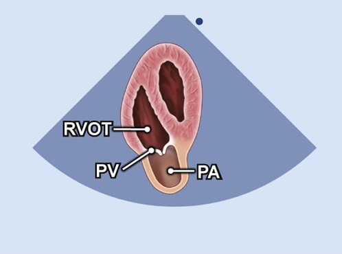
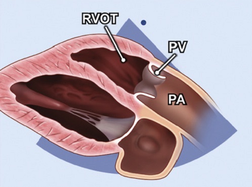
Right ventricular outflow tract view (fetal echocardiogram) | Radiology  Reference Article | Radiopaedia.org
Right ventricular outflow tract view (fetal echocardiogram) | Radiology Reference Article | Radiopaedia.org

5A. Parasternal long axis view of the right ventricular outflow tract  (normal) - YouTube
5A. Parasternal long axis view of the right ventricular outflow tract (normal) - YouTube

Cardiac STRUCTURES! (RV inflow and outflow view) - Echocardiography! -  YouTube
Cardiac STRUCTURES! (RV inflow and outflow view) - Echocardiography! - YouTube

Echocardia - Wiki
Echocardia - Wiki

A5C focus on RVOT (Echo) - TECHmED

Right Ventricular Outflow Tract (RVOT) Changes in Children with an Atrial  Septal Defect: Focus on RVOT Velocity Time Integral, RVOT Diameter, and RVOT  Systolic Excursion - Koestenberger - 2016 - Echocardiography - Wiley Online  Library
Right Ventricular Outflow Tract (RVOT) Changes in Children with an Atrial Septal Defect: Focus on RVOT Velocity Time Integral, RVOT Diameter, and RVOT Systolic Excursion - Koestenberger - 2016 - Echocardiography - Wiley Online Library

17: Anomalies of the Right Ventricular Outflow Tract and Pulmonary Valve |  Veterian Key
17: Anomalies of the Right Ventricular Outflow Tract and Pulmonary Valve | Veterian Key

Non-Circular Shape of Right Ventricular Outflow Tract | Circulation:  Cardiovascular Imaging
Non-Circular Shape of Right Ventricular Outflow Tract | Circulation: Cardiovascular Imaging

JCM | Free Full-Text | A Novel Diagnostic Score Integrating Atrial  Dimensions to Differentiate between the Athlete's Heart and Arrhythmogenic  Right Ventricular Cardiomyopathy
JCM | Free Full-Text | A Novel Diagnostic Score Integrating Atrial Dimensions to Differentiate between the Athlete's Heart and Arrhythmogenic Right Ventricular Cardiomyopathy

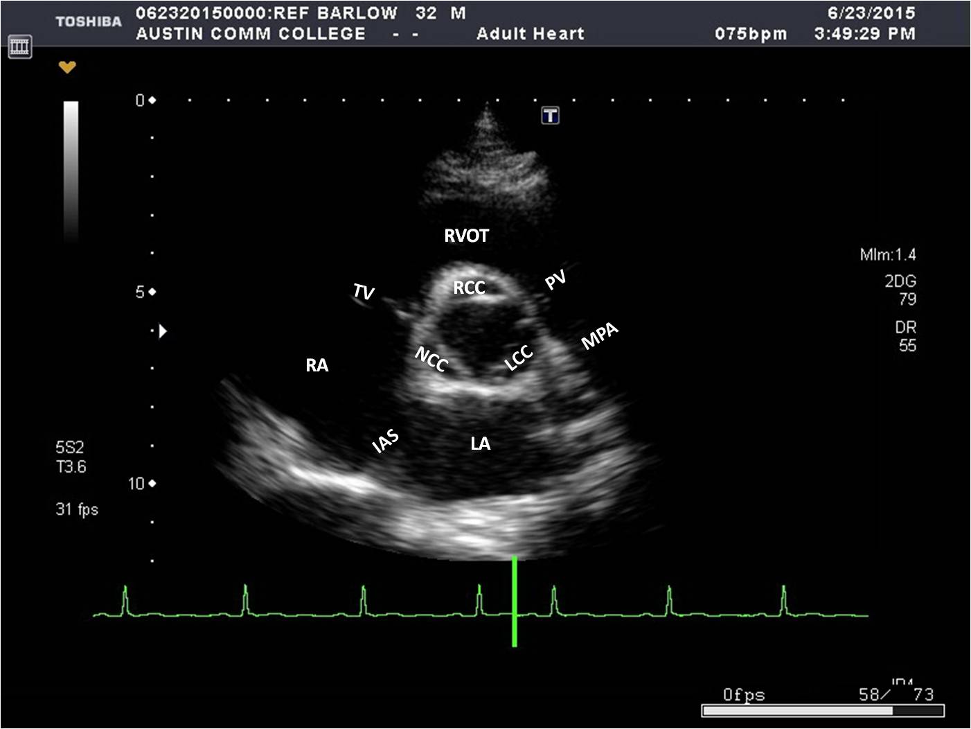
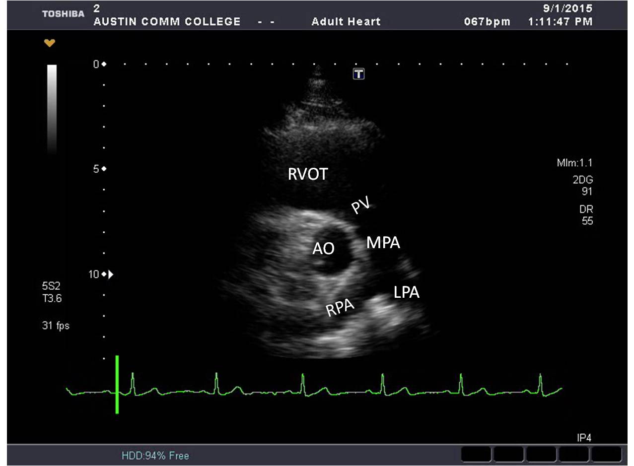
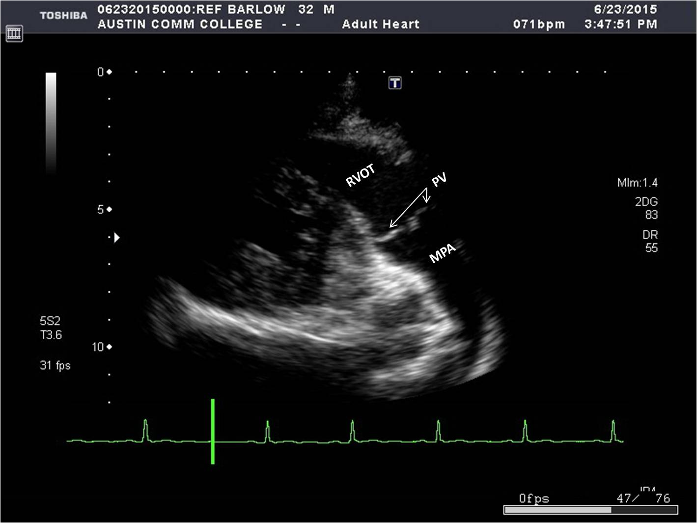
Parasternal Window | Sonography Resources

A case of subvalvular pulmonary stenosis differentiated from a  double-chambered right ventricle by transesophageal echocardiography:  importance of detecting the pulmonary valve | SpringerLink
A case of subvalvular pulmonary stenosis differentiated from a double-chambered right ventricle by transesophageal echocardiography: importance of detecting the pulmonary valve | SpringerLink

RVOT View TEE | Cardiac sonography, Diagnostic medical sonography,  Diagnostic medical sonography student
RVOT View TEE | Cardiac sonography, Diagnostic medical sonography, Diagnostic medical sonography student

PLAX RV outflow (Echo) - TECHmED

7 Tips to Implementing Right Ventricle Quantification Cardioserv

Right ventricular outflow tract view. A, The transducer is held over... |  Download Scientific Diagram
Right ventricular outflow tract view. A, The transducer is held over... | Download Scientific Diagram

Parasternal Window | Sonography Resources

Guidelines for the Echocardiographic Assessment of the Right Heart in  Adults: A Report from the American Society of Echocardiogr
Guidelines for the Echocardiographic Assessment of the Right Heart in Adults: A Report from the American Society of Echocardiogr

3.3.2 Right ventricular size | 123 Sonography

Two-dimensional view of right ventricular outflow tract at end-diastole...  | Download Scientific Diagram
Two-dimensional view of right ventricular outflow tract at end-diastole... | Download Scientific Diagram

Parasternal Window | Sonography Resources

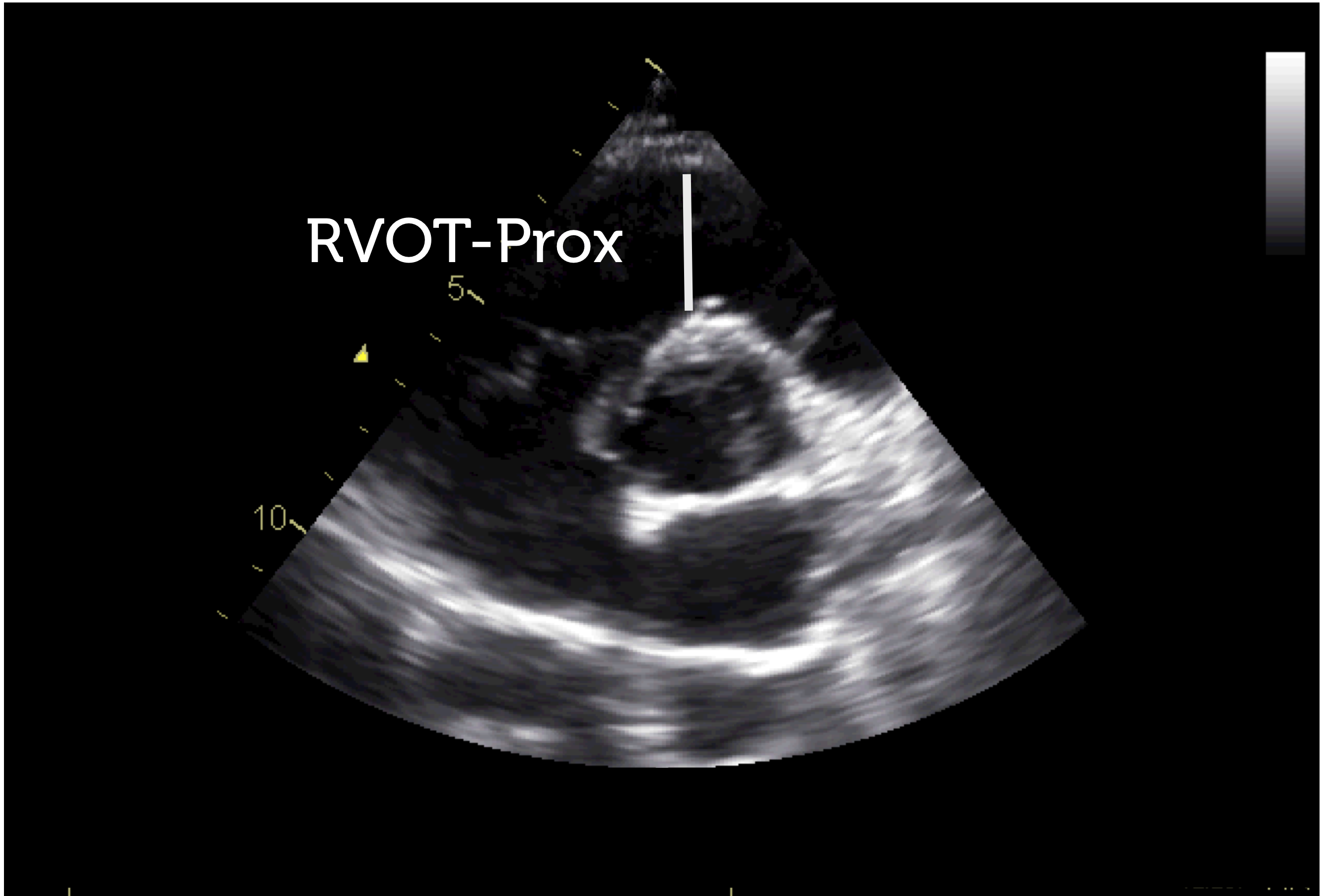
Echo parasternal views RVOT measurements